
Fig. S4
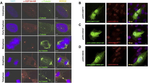
Subcellular Localization of CEP164 and SDCCAG8 by Immunofluorescence (A) CEP164 colocalizes with the mother centriole (labeled with γ-tubulin), the mitotic spindle poles (actetylated tubulin), and the midbody throughout the cell cycle in hTERT-RPE cells. Whereas in the centriole-engaged state (upper panel) γ-tubulin antibody labels both centrioles, the α-CEP164 antibody only labels one centriole (the mother centriole) in hTERT cells. Immunofluorescence of endogenous CEP164 was performed in hTERT cells. Cells were fixed (4% PFA), permeabilized (0.1% SDS) and immuno-stained with antibody anti-CEP164-NR (red) and costained with anti-γ-tubulin or anti-acetylated tubulin antibodies (green). DAPI was used to label DNA (Blue). Scale bars, 2.5 μm. (B)–(D) Upon immunofluorescence (IF) in hTERT-RPE cells antibody α-SDCCAG8-CG recognizes nuclear foci that are absent upon transient SDCCAG8/NPHP10 knockdown. Left panels show transfected hTERT-RPE cells, middle panels show IF with α-SDCCAG8-CG, right panels show merge of left and middle panels. Antibody α-SDCCAG8-CG recognizes nuclear foci in hTERT-RPE cells (middle and right panels). (B) hTERT cells transiently transfected with GFP-labeled negative control shRNA construct exhibit nuclear foci upon IF with α-SDCCAG8-CG (arrow heads in right panel), whereas in cells transfected with GFP-labeled SDCCAG8 shRNA knockdown constructs pGIPZ-259547 (C) or pGIPZ-90858 (D) nuclear foci are absent (asterisks in right panels of (C) and (D), demonstrating specificity of the nuclear foci signal detected by α-SDCCAG8-CG. Scale bars, 5 μm. See also Figure 4. |
Reprinted from Cell, 150(3), Chaki, M., Airik, R., Ghosh, A.K., Giles, R.H., Chen, R., Slaats, G.G., Wang, H., Hurd, T.W., Zhou, W., Cluckey, A., Gee, H.Y., Ramaswami, G., Hong, C.J., Hamilton, B.A., Cervenka, I., Ganji, R.S., Bryja, V., Arts, H.H., van Reeuwijk, J., Oud, M.M., Letteboer, S.J., Roepman, R., Husson, H., Ibraghimov-Beskrovnaya, O., Yasunaga, T., Walz, G., Eley, L., Sayer, J.A., Schermer, B., Liebau, M.C., Benzing, T., Le Corre, S., Drummond, I., Janssen, S., Allen, S.J., Natarajan, S., O'Toole, J.F., Attanasio, M., Saunier, S., Antignac, C., Koenekoop, R.K., Ren, H., Lopez, I., Nayir, A., Stoetzel, C., Dollfus, H., Massoudi, R., Gleeson, J.G., Andreoli, S.P., Doherty, D.G., Lindstrad, A., Golzio, C., Katsanis, N., Pape, L., Abboud, E.B., Al-Rajhi, A.A., Lewis, R.A., Omran, H., Lee, E.Y., Wang, S., Sekiguchi, J.M., Saunders, R., Johnson, C.A., Garner, E., Vanselow, K., Andersen, J.S., Shlomai, J., Nurnberg, G., Nurnberg, P., Levy, S., Smogorzewska, A., Otto, E.A., and Hildebrandt, F., Exome Capture Reveals ZNF423 and CEP164 Mutations, Linking Renal Ciliopathies to DNA Damage Response Signaling, 533-548, Copyright (2012) with permission from Elsevier. Full text @ Cell