FIGURE

Fig. S2

ID
ZDB-FIG-120727-9
Publication
Danesin et al., 2009 - Integration of telencephalic Wnt and hedgehog signaling center activities by Foxg1
Other Figures
All Figure Page
Back to All Figure Page
Fig. S2

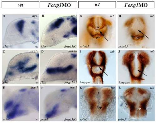
Neuronal Differentiation Is Affected in the Telencephalon of foxg1 Morphant
(A–F) Lateral views, anterior to the left, G-L: frontal views, dorsal top. Developmental stages are indicated at bottom left of each picture. (A) ngn1 expression is detected in the dorsal telencephalic neurons in wt embryos and its expression expands ventrally in foxg1 morphants (B). Ventral neurons detected by expression of zash1a (C) or islet1 (E) are absent in foxg1 morphants (D and F, respectively).
Anti-tubulin immunostaining at later stages show that the anterior commissure (arrows in G and I) is completely absent in foxg1 knockdown embryos (H and J), while the postoptic commissure remains unaffected. Hu immunostaining labelling neurons nuclei in foxg1 morphants (L) in comparison with wt (K). t, telencephalon ; hyp, hypothalamus; d, diencephalon ; ac, anterior commissure ; poc, post-optic commissure.

Expression Data

Expression Detail
Antibody Labeling
Phenotype Data

Phenotype Detail
Acknowledgments
This image is the copyrighted work of the attributed author or publisher, and ZFIN has permission only to display this image to its users. Additional permissions should be obtained from the applicable author or publisher of the image.

Reprinted from Developmental Cell, 16(4), Danesin, C., Peres, J.N., Johansson, M., Snowden, V., Cording, A., Papalopulu, N., and Houart, C., Integration of telencephalic Wnt and hedgehog signaling center activities by Foxg1, 576-587, Copyright (2009) with permission from Elsevier. Full text @ Dev. Cell