
Fig. 4
- ID
- ZDB-FIG-120718-23
- Publication
- Cibrián Uhalte et al., 2012 - In vivo conditions to identify prkci phosphorylation targets using the analog-sensitive kinase method in zebrafish
- Other Figures
- All Figure Page
- Back to All Figure Page
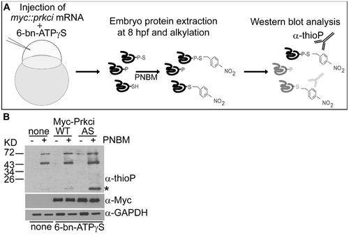
Thiophosphorylation of substrate proteins by PrkciI316 in the zebrafish embryo. (A) Schematic diagram of the in vivo labeling method for the selective labeling of PrkciI316A substrates during zebrafish development. (B) In vivo thiophosphorylation in zebrafish embryos injected at the one-cell stage with 200 μM N6-benzyl-ATPγS (6-bn-ATPγS) and mRNA encoding either PrkciWT or PrkciI316A (AS). Western blot analysis with rabbit monoclonal anti-thiophosphoester (α-thioP) C51-8 antibody (Epitomics) of 80% epiboly (6–8hpf) samples alkylated with 2.5 mM PNBM reveals a selectively labeled protein in the PrkciI316A (AS) sample (asterisk). |