
Fig. 7
- ID
- ZDB-FIG-120710-74
- Publication
- Hörndli C.S. et al., 2012 - Sonic hedgehog is indirectly required for intraretinal axon pathfinding by regulating chemokine expression in the optic stalk
- Other Figures
- All Figure Page
- Back to All Figure Page
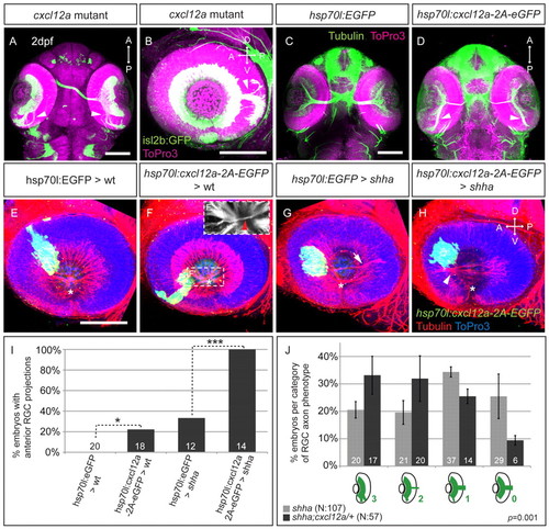
Cxcl12a acts as an RGC axonal attractant in wt and shha and interacts genetically with the Hh pathway in zebrafish. (A-H) Maximum-intensity projections of ventral (A,C,D) and lateral (B,E-H) views at 2 dpf. (A,B) Cxcl12a mutants exhibit intraretinal axon guidance errors (arrowheads). Isl2b:GFP (green); nuclei, ToPro3 (magenta). (C) Normal axonal projections in hsp70l:EGFP embryos after heatshock. (D) Global Cxcl12a-2A-EGFP overexpression induces intraretinal axon guidance errors (arrowheads). α-tubulin (pseudocolored green); nuclei, ToPro3 (magenta), EGFP not shown. (E-H) Cxcl12a-expressing cells attract RGC axons in wt and shha embryos. Transplanted EGFP- or cxcl12a-2A-EGFP-expressing cells (green), α-tubulin (red); nuclei, ToPro3 (blue). (E) Anterior EGFP-expressing cells in wt embryos do not affect RGC outgrowth. (F) Anterior projections in wt embryos with anterior Cxcl12a-expressing cells. (F′) Substack of boxed region in F with misguided axons (red arrowhead). (G) Rare anterior projections in shha embryos with EGFP-expressing cells. (H) Shha embryos always show anterior projections with anterior Cxcl12a-expressing cells. Optic disc, asterisk. D, dorsal; V, ventral; A, anterior; P, posterior. Scale bars: 100 μm. (I) Percentage of host embryos with anterior RGC projections. Number of embryos shown at base of bars. *P<0.05, ***P<0.001, Fisher′s exact test. (J) Analysis of genetic interaction between shha and cxcl12a. Percentage of embryos per category (0-3, illustrated below graph) of RGC axon projection phenotype in shha (light gray) and shha;cxcl12a/+ (dark gray). Error bars represent s.e.m. n=3 experiments. Mann-Whitney U test, P=0.00103, of embryos ranked in four categories in shha (n=107) and shha;cxcl12a/+ (n=57) populations. |