
Fig. 6
- ID
- ZDB-FIG-120508-4
- Publication
- Stooke-Vaughan et al., 2012 - The role of hair cells, cilia and ciliary motility in otolith formation in the zebrafish otic vesicle
- Other Figures
- All Figure Page
- Back to All Figure Page
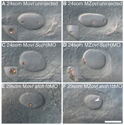
The effect of ectopic hair cells and loss of hair cells on otolith formation in the absence of cilia. All panels show left ear with anterior to the left. (A) 24S uninjected Movl control embryo; inset shows anterior otolith on tether cilia. (B) 24S uninjected MZovl mutant embryo; inset shows small anterior otolith tethered to the hair cells’ apical surface at the anterior OV pole. (C) 24S Movl control embryo injected with Su(H)1+2MO; inset shows otolith bound to several ectopic tether cilia. (D) 24S MZovl mutant embryo injected with Su(H)1+2MO; inset shows otolith precursor particles tethered to the apical surface of the ectopic hair cells. (E) 29S Movl control embryo injected with atoh1bMO; one untethered otolith is present in the ear. (F) 29S MZovl mutant embryo injected with atoh1bMO; one untethered otolith (slightly out of focal plane) is present in the ear (arrowhead). The ovl genotype was confirmed for all samples by genotyping (data not shown). Scale bar: 40 μm for A-F. |