
Fig. 2
- ID
- ZDB-FIG-120507-12
- Publication
- Stooke-Vaughan et al., 2012 - The role of hair cells, cilia and ciliary motility in otolith formation in the zebrafish otic vesicle
- Other Figures
- All Figure Page
- Back to All Figure Page
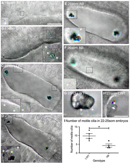
Ciliary motility in the developing wild-type zebrafish otic vesicle. All panels are dorsolateral views of the left ear, with anterior to the right, and are merged time-to-colour composites of six consecutive frames of a high-speed movie. Grey scale shows lack of movement; colour indicates movement. White asterisks mark motile cilia. (A) 18S AB strain wild-type embryo with one motile cilium present on the medial wall (shown in inset). See supplementary material Movie 1. (B) 19S LWT wild-type embryo. Inset shows motile cilia next to the anterior otolith and movement of the anterior otolith. See supplementary material Movie 2. (C) 22S LWT wild-type embryo. Insets: motile cilia present on the medial wall of the OV (two in the left hand inset, one in the right hand inset). See supplementary material Movie 3. (D) 23S LWT wild-type embryo. The posterior otolith is shown on two immotile tether cilia (grey); three motile cilia are nearby (coloured). See supplementary material Movie 4. (E) 26S AB wild-type embryo. Inset: motile cilia next to posterior otolith. See supplementary material Movie 5. (F) Different focal plane of the embryo shown in E. One motile cilium was present on the medial wall of the OV (inset) that rotated first one way and then the other. See supplementary material Movie 6. (G) 25 hpf LWT wild-type embryo. The otolith sits on two to three tether cilia; two short, motile cilia remain directly underneath. See supplementary material Movie 7. (H) Very rarely, one or a few otolith precursor particles were bound to a motile cilium (asterisk; 22S LWT wild-type embryo). See supplementary material Movie 8. (I) Number of motile cilia found per OV in AB and LWT wild-type strains. Bars represent mean ± s.e.m.. Scale bars: 20 μm in A, for A-F; 10 μm in H, for G,H and insets in A-F. |