
Fig. 4
- ID
- ZDB-FIG-120507-14
- Publication
- Stooke-Vaughan et al., 2012 - The role of hair cells, cilia and ciliary motility in otolith formation in the zebrafish otic vesicle
- Other Figures
- All Figure Page
- Back to All Figure Page
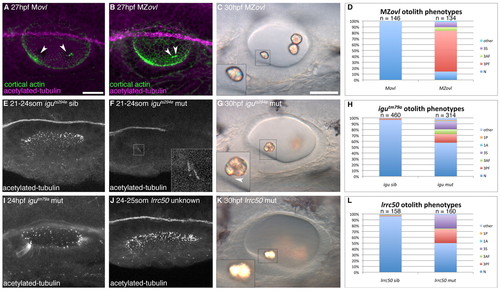
Formation of otoliths in the absence of cilia. (A) Anti-acetylated tubulin and FITC-phalloidin stain showing tether cilia in an Movl embryo (arrowheads; magenta). (B) MZovl mutant ears lack all cilia, but stereociliary bundles are still present (arrowheads; green). (C) Imperfect otoliths form in the correct position in MZovl. Inset: otolith resting directly on a stereociliary bundle. (D) Graph of otolith defects in MZovl embryos. (E) Confocal stack showing cilia in a phenotypically wild-type iguts294e sibling embryo. (F) Anterior kinocilia present in an iguts294e mutant (inset). (G) The iguts294e mutant lacks most cilia but still has some tether cilia (inset, arrowhead). (H) Graph of otolith defects in igutm79a. (I) The igutm79a mutant allele has more otic cilia than the iguts294e allele, but fewer than in wild-type embryos. (J) Representative confocal stack of an ear from an lrrc50 cross. It was not possible to distinguish lrrc50 mutant from sibling embryos at <24 hpf (n=15 ears). (K) DIC image of the ear of an lrrc50 mutant. (L) Graph of lrrc50 mutant otolith phenotypes. N, normal (two otoliths, in correct positions); 1A, one anterior otolith; 1P, one posterior otolith; 3S, three separate otoliths; 3AF, three otoliths, with two anterior otoliths fused; 3PF, three otoliths, with two posterior otoliths fused; other, other otolith defects. Scale bars: 25 μm in A, for A,B,E,F,I,J; 30 μm in C, for C,G,K. |