
FIGURE
Fig. S2
Fig. S2
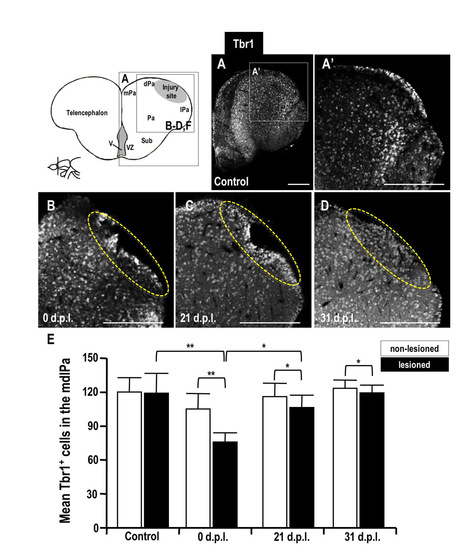
Distribution of Tbr1-positive cells in the adult zebrafish telencephalon. (A-D) Immunodetection of Tbr1 protein in coronal brain sections (dorsal up) of the lesioned adult zebrafish brain: (A, A′) control; (B) 0 d.p.l.; (C) 21 d.p.l.; (D) 31 d.p.l.. Yellow dotted circles indicate the injury site. (E) Histogram showing the Tbr1-positive cell counts in the medial-dorsal-lateral domain of the telencephalic pallium (mdlPa) in the injured brains of adult wild-type zebrafish. Scale bars: 100 μm. Abbreviations: see Fig. 2. |
Expression Data
Expression Detail
Antibody Labeling
Phenotype Data
Phenotype Detail
Acknowledgments
This image is the copyrighted work of the attributed author or publisher, and
ZFIN has permission only to display this image to its users.
Additional permissions should be obtained from the applicable author or publisher of the image.
Full text @ Dis. Model. Mech.