
Fig. 1
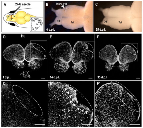
The healing process in the adult zebrafish telencephalon. (A) A zebrafish model of adult telencephalon lesions. A lesion was formed by inserting a needle into the dorsolateral domain of one hemisphere of the telencephalon. (A′) The inset is an illustration of the coronal hemisphere section obtained by a cut at the red line. Tel, telencephalon; mPa, medial pallium; dPa, dorsal pallium; Pa, pallium; lPa, lateral pallium; Sub, subpallium. (B,C) Dorsal views of an injured adult zebrafish brain at 0 days post-lesion (dpl) (B) and 35 dpl (C). Note the reduction of the lesion at 35 dpl. (D–F) Hu protein detected in coronal brain sections at the level indicated in the illustration in panel D (dorsal up) of zebrafish at 1 (D), 14 (E) and 35 (F) dpl. Higher magnifications of the boxed regions in panels D–F are shown in D2–F2, respectively. Arrows indicate accumulated Hu-positive cells at the injury site, which is circled in white. Scale bars: 100 μm. |