|
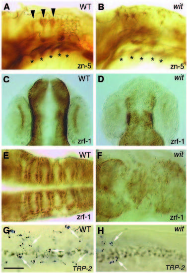
Some cell types are decreased in witta52b. C and D are rostral to the top. A, B are lateral views; C-H are dorsal views. Wildtype (A) and mutant (B) embryos labeled with zn-5 mAb at 36 hpf. The dorsal hindbrain neurons (arrowheads; Trevarrow et al., 1990) are decreased in mutant embryos. The pharyngeal endoderm (asterisks; Schilling and Kimmel, 1994) is not affected. The pharyngeal endoderm in B is out of focus. Wildtype (C,E) and mutant (D,F) embryos labeled with zrf-1 mAb. C,D are at 24 hpf; E,F are at 38 hpf. Radial glial fibers in the retina (Westerfield, 1994) and hindbrain (Trevarrow et al., 1990) are decreased. TRP-2 expression pattern of wild-type (G) and mutant (H) embryos in the anterior trunk at 24 hpf. The labeled melanophores (arrows) are decreased in H. Bar, 125 µm (A-D); 100 µm (E-H).
|