
Fig. 5
- ID
- ZDB-FIG-120216-94
- Publication
- Goonesinghe et al., 2012 - Desmosomal cadherins in zebrafish epiboly and gastrulation
- Other Figures
- All Figure Page
- Back to All Figure Page
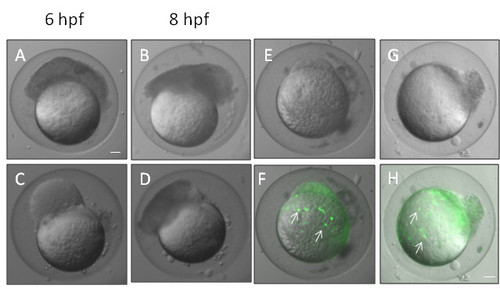
Knockdown of desmosomal cadherins causes epiboly defects. A and C show morphants at 6 hpf whose blastomeres became detached and failed to migrate towards the vegetal pole. The blastomeres of these embryos still continued to divide and seemed to undergo movements resembling gastrulation movements (B, D). These images are representative of similar phenotypes obtain by knockdown of both zfDsc1 and zfDsg2. E, F and G, H show embryos that were injected with Sytox to stain the nuclei of the YSL (white arrows in F, H). The DIC images (E, G) show that the blastomeres had become detached while the merged DIC and fluorescence images (F, H) show that the YSL had continued epiboly movement toward the vegetal pole. (Embryos such as those shown in this figure did not survive beyond about 10 hpf and are recorded as dead in Figure 3.) E, F were obtained by knockdown of Dsc1 and G, H by knockdown of Dsg2. Scale bars = 100 μm. |
| Fish: | |
|---|---|
| Knockdown Reagents: | |
| Observed In: | |
| Stage: | Shield |