
Fig. 8
- ID
- ZDB-FIG-120216-97
- Publication
- Goonesinghe et al., 2012 - Desmosomal cadherins in zebrafish epiboly and gastrulation
- Other Figures
- All Figure Page
- Back to All Figure Page
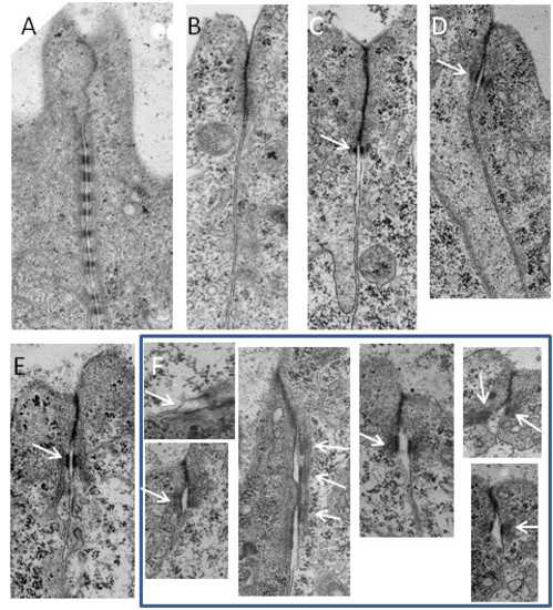
Knockdown of desmosomal cadherins affects desmosome structure. A. Apical junctional complex between cells of the EL of wild type embryo at 24 hpf showing the tight junction/adherens junction region (tj, aj) and the extensive array of desmosomes (d). At epiboly (B) the tight junction/adherens junction was present but no desmosomes were found. By the shield stages (C) fully formed desmosomes (arrow) were present forming the lowest component of the junctional complex in wild type embryos. D. A single fully formed desmosome (arrow) found in a morpholino injected embryo that reached the shield stage. At the 8 somite stage (E) desmosomes (arrow) were present at the lower aspect of the junctional complex in wild type embryos. F. A representative sample of abnormal desmosomes (arrows) form morpholino injected embryos that reached the 8 somite stage. Scale bars: A = 1 μm; B-F = 0.5 μm. |
| Fish: | |
|---|---|
| Knockdown Reagent: | |
| Observed In: | |
| Stage Range: | 5-9 somites to Prim-5 |