
Fig. 2
- ID
- ZDB-FIG-120216-38
- Publication
- Asakawa et al., 2012 - An mnr2b/hlxb9lb enhancer trap line that labels spinal and abducens motor neurons in zebrafish
- Other Figures
- All Figure Page
- Back to All Figure Page
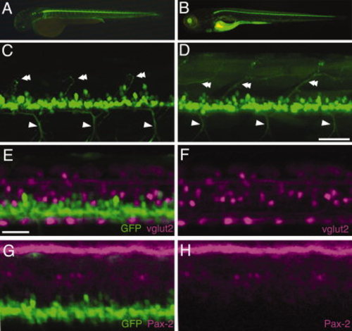
Green fluorescent protein (GFP) expression in the HGj4A embryos and larvae. A,B: The lateral view of the HGj4A embryo at 48 hpf (A) and larva at 120 hours postfertilization (hpf; B). C,D: The lateral view of the spinal cord in HGj4A embryo at 48 hpf (C) and larva at 120 hpf (D). The arrowheads and double arrowheads indicate the motor axons extending ventrally and dorsally, respectively. E,F: The single section the spinal cord of the HGj4A;Tg(vglut2a: loxPDsRed-GFP) double transgenic embryo at 48 hpf. In E, the GFP signal was merged with the red fluorescent protein (RFP) signal (vlut2, F). G,H: The single section of the spinal cord of HGj4A embryo at 48 hpf, stained with anti-Pax-2 antibody (magenta). In G, the GFP signal was merged with the Pax-2 signal (magenta, H). Scale bars = 50 μm in C,D, 30 μm in E–H. |