
Fig. 1
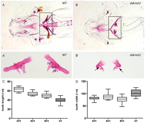
ext2-/- mutant displays severe tooth phenotype. Ventral view of alizarin-red-stained craniofacial skeleton and teeth at 6 dpf (A, B) and dissected and flat mounted 5th pharyngeal arches with teeth (A2, B2) reveals the presence on each pharyngeal arch of 3 teeth in siblings (A, A2) and only one misshapen tooth in ext2-/- larvae (B, B2). Note that the rod shaped branchial arch 5 to which the teeth attach is also ossified. Arrows point incomplete ossification of the mutant tooth. Tooth phenotype consisting of one misshapen tooth was observed in all (n>500) analysed ext2-/- embryos whereas heterozygote fish were indistinguishable from WT. Tooth lengths varies between 3V1, 4V1 and 5V1 in siblings (P<0.003). Each of those teeth was significantly longer then dak-tooth (P<0.0001) (C). Tooth widths of 3V1 and 5V1 were similar between siblings, and both were significantly narrower than 4V1 (D). ext2-/--tooth was significantly broader than any of the siblings teeth (3V1, P<0.0001; 4V1, P = 0.023 and 5V1, P = 0.0001) (D). White boxes, siblings; grey boxes, homozygote mutant. Scale bar = 0.1 mm. |
| Fish: | |
|---|---|
| Observed In: | |
| Stage: | Day 6 |