
Fig. 7
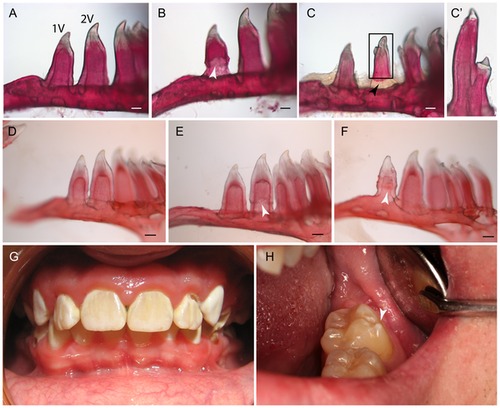
Dental defects are present in 20% of adult ext2+/- mutant fish. Lateral view of two ventral teeth stained with Alizarin red. In most cases, WT-like teeth were present (A, D). However, on few occasions we also observed: enamel malformation (B, E, F) or misshapen crowns (C, C2). Teeth start to calcify from the tip toward the base; hence the lack of staining at the base of 2V is most likely reflects uncompleted ossification of a recent replaced tooth – see black arrowhead (C). Teeth from MO patients (G, H). Note extra buckle in H (arrow head) which resembles split crown observed in ext2-/- fish. C2 is a higher magnification of C. White arrows indicate lesions. Scale bars correspond to 0.1 mm. |
| Fish: | |
|---|---|
| Observed In: | |
| Stage: | Adult |