
Fig. 6
- ID
- ZDB-FIG-120201-31
- Publication
- Chen et al., 2011 - Role of zebrafish lbx2 in embryonic lateral line development
- Other Figures
- All Figure Page
- Back to All Figure Page
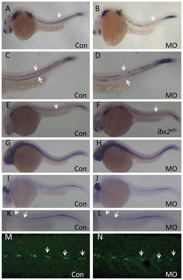
Abrogation of zebrafish lbx2 results in a defective sdf1a expression pattern in the horizontal myoseptum. (A, B) Compared with control embryos (A), a weak and discontinuous sdf1a expression pattern is observed in the horizontal myoseptum of lbx2 morphants at 24 hpf (B; lateral view). (C, D) Magnified views of the expression patterns shown in A (C) and B (D). (E, F) Compared with gfp mRNA-injected control embryos (E), injection of lbx2eh- mRNA resulted in a defective sdf1a expression at 24 hpf (F). (G, H) Similar expression pattern of tenascin C, a marker of the horizontal myoseptum, in control embryos (G) and lbx2 morphants (H) at 30 hpf. (I, J) Analysis of eng2a positive muscle pioneer cells in control embryos (I) and lbx2 morphants (J) at 30 hpf. (K, L) Magnified views of the expression patterns shown in I (K) and J (L), showing decreased numbers of eng2a positive muscle pioneer cells in lbx2 morphants (L) compared to control embryos (K). (M, N) Compared with control embryos (M), the numbers of 4D9 positive muscle pioneer cells was slightly reduced in lbx2 morphants (N) at 30 hpf. White arrows indicate the hybridization or immunostaining signals. All images are lateral views. |