
FIGURE
Fig. S2
- ID
- ZDB-FIG-120201-25
- Publication
- Chen et al., 2011 - Role of zebrafish lbx2 in embryonic lateral line development
- Other Figures
- All Figure Page
- Back to All Figure Page
Fig. S2
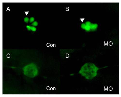
PLL cells in the newly deposited neuromasts of lbx2 morphants appear normal. (A–B) Hair cells of PLL neuromasts labeled with GFP in the SqET4 transgenic zebrafish line. The pattern and numbers of PLL hair cells in newly deposited neuromasts was similar in control embryos (A) and lbx2 morphants (B) at 48 hpf. (C, D) Fluorescence images of SqET10 embryos indicating that the supporting cells and lateral line nerve in newly deposited neuromasts of embryos injected with control MO (C) or lbx2 MO (D) are similar at 48 hpf. The white arrowhead indicates HCs in deposited neuromasts. |
Expression Data
Expression Detail
Antibody Labeling
Phenotype Data
Phenotype Detail
Acknowledgments
This image is the copyrighted work of the attributed author or publisher, and
ZFIN has permission only to display this image to its users.
Additional permissions should be obtained from the applicable author or publisher of the image.
Full text @ PLoS One