
Fig. 6
- ID
- ZDB-FIG-120118-18
- Publication
- Jung et al., 2011 - Aberrant hedgehog ligands induce progressive pancreatic fibrosis by paracrine activation of myofibroblasts and ductular cells in transgenic zebrafish
- Other Figures
- All Figure Page
- Back to All Figure Page
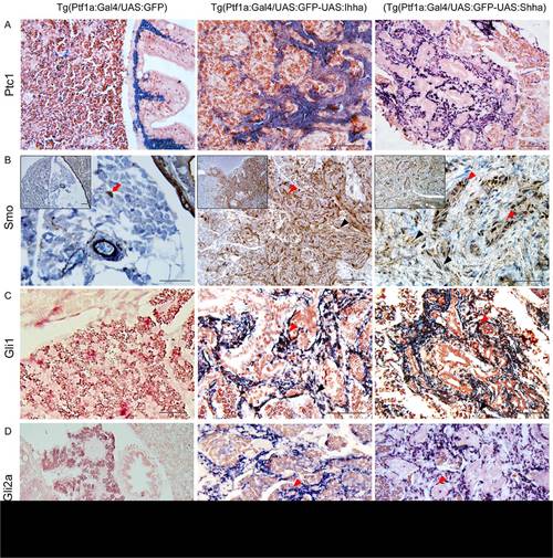
Expression of the downstream components of Hh signaling at 6 month-old zebrafish pancreas. (A) ISH for Ptc1. In control Ptc1 is expressed in the muscle layer of bowel and pancreatic duct. In the Hh-expressing pancreas, both proliferating myofibroblasts and ductular cells express Ptc1. (B) IHC for Smo reveals strong expression in a wide area of fibrosis. Both myofibroblasts and ductular cells are reactive to Smo. Likely to α-SMA immunostaining, Smo-reactive cells (red arrow) are occasionally noted in the parenchyma of the control pancreas. Inlets are 200× images. (C, D) ISH for Gli1 and Gli2a. Whereas, the control pancreas reveals a negligible degree of Gli1 and Gli2a expression, activated myofibroblasts and ductular cells express Gli1 and Gli2a. (B–D)Black arrowheads, myofibroblasts; Red arrowheads, ductular cells. If not specified, microscopic images are 400×. Bars, 50 μm. |