
Fig. 3
- ID
- ZDB-FIG-120118-15
- Publication
- Jung et al., 2011 - Aberrant hedgehog ligands induce progressive pancreatic fibrosis by paracrine activation of myofibroblasts and ductular cells in transgenic zebrafish
- Other Figures
- All Figure Page
- Back to All Figure Page
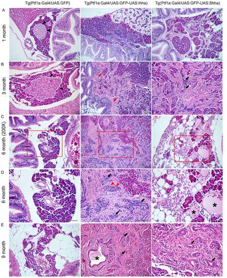
Histopathologic findings showing progressive pancreatic fibrosis. (A) Progressive pancreatic fibrosis starts at as early as 1-month old in Hh-expressing transgenic zebrafish. In non-fibrotic area, Individual morphology of the pancreatic acini and acinar cells is not unusual. (B) A principal islet is seen in control, which is well-circumscribed by acinar cells (black arrowheads). In Hh-secreting lines, accumulation of fibrosis results in the destruction of the morphologic architecture, which is prominent even at 3 months. Fibrotic bands are contiguous from the bowel wall forming adhesion between the bowel and the pancreas (red arrows), suggesting recruitment of myofibroblasts from the muscle layer of the bowel. Along with fibrosis, an increasing number of ductular structure appears within fibrotic area at 3 months of age (black arrows). (C, D) The pancreas at 6-months old. (D) An enlarged view of the red box in (C). Contrary to the islet of control in B, some islets of the Hh-expressing pancreas are completely encircled by fibrosis (red arrowheads), which is typical finding in chronic pancreatitis of human. The number of ductular structure further increased (black arrows). Occasionally, acute pancreatitis-like changes are noted, showing the infiltration of inflammatory cells and cystic space filled with mucinous material (asterisks). (E) The exocrine pancreas of 9 month-old zebrafish shows more accumulation of fibrosis and ductular structures (black arrows). At center image, a large pancreatic duct (asterisk) is seen, being surrounded by fibrosis and ductular structures. If not specified, microscopic images are 400×.Bars, 50 μm. |