
Fig. 6
- ID
- ZDB-FIG-120105-2
- Publication
- Song et al., 2011 - The role of stat1b in zebrafish hematopoiesis
- Other Figures
- All Figure Page
- Back to All Figure Page
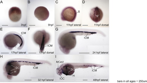
Whole mount RNA in situ hybridization shows specific expression pattern of stat1b in zebrafish hematopoietic regions. A, B. 3 and 9 hpf embryos, respectively, did not show specific expression of stat1b. C and D. At 11 hpf (2 somites), stat1b was expressed in stripes flanking the paraxial mesoderm, shown in lateral and dorsal views, respectively. E and F. At 17 hpf (18 somites), stat1b was expressed in the intermediate cell mass, shown in lateral and dorsal views, respectively. G. By 24 hpf, stat1b expression stripes had merged centrally. H. At 32 hpf, the stat1b expression compartment had spread from the intermediate cell mass to the heart and ducts of Cuvier on the yolk. I. At 48 hpf, the expression of stat1b continued in hematopoietic regions and veins. Abbreviations: dc, ducts of Cuvier; h, heart; icm, intermediate cell mass; IOC, inner optic circle; MCeV, middle cerebral vein ( Isogai et al., 2001); s, stripes flanking the paraxial mesoderm. |
| Gene: | |
|---|---|
| Fish: | |
| Anatomical Terms: | |
| Stage Range: | 1k-cell to Long-pec |
Reprinted from Mechanisms of Development, 128(7-10), Song, H., Yan, Y.L., Titus, T., He, X., and Postlethwait, J.H., The role of stat1b in zebrafish hematopoiesis, 442-56, Copyright (2011) with permission from Elsevier. Full text @ Mech. Dev.