
Fig. 7
- ID
- ZDB-FIG-111115-7
- Publication
- Lister et al., 2011 - Embryonic expression of zebrafish MiT family genes tfe3b, tfeb, and tfec
- Other Figures
- All Figure Page
- Back to All Figure Page
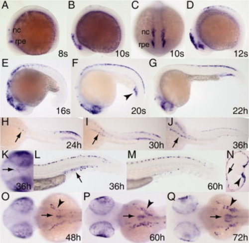
Expression pattern of tfec during embryogenesis. Whole mount RNA in situ hybridization was performed on embryos at the stages indicated. A–F: Expression begins in the posterior eye region/presumptive retinal pigment epithelium and a few neural crest cells at the eight-somite stage (A) and expands during somitogenesis (B–F). H–J,O–Q: The tfec-expressing cells populate bilateral patches over the yolk beginning at 24 hpf (arrows, H–J), increasing in number and extent through 72 hours postfertilization (hpf; arrowheads, O–Q), and are also found on top of the head (arrow, K) and dorso- and ventromedially in the trunk and tail (L,M), all positions characteristic of iridophores. F,L,M: Expression in the intermediate cell mass is seen by the 20-somite stage (arrowhead, F), has weakened by 36 hpf (arrowhead, L) and is gone by 60 hpf (M). Expression in the retinal pigment epithelium (arrow) and ciliary margin (arrowhead) is observed in transverse section of the eye at 60 hpf (N, dorsal is to the top). O–Q: Beginning at 48 hpf, expression is observed in the developing swim bladder (arrows). Lateral views A,B,D–G,L,M; frontal view, C; dorsolateral views, H–J; dorsal views K,O–Q. nc, neural crest; rpe, (presumptive) retinal pigment epithelium. |
| Gene: | |
|---|---|
| Fish: | |
| Anatomical Terms: | |
| Stage Range: | 5-9 somites to Protruding-mouth |