
Fig. 7
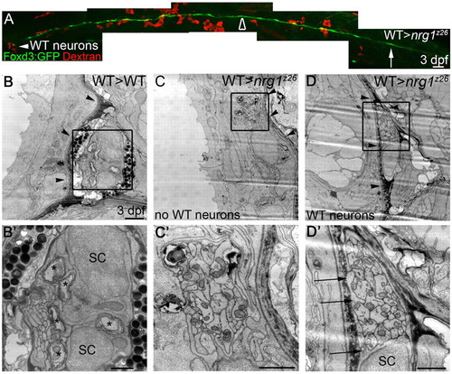
Schwann cells can migrate along nrg1z26 mutant axons in the presence of wild-type neurons. (A) Lateral view of 3 dpf genetic chimera generated by transplanting Dextran-labeled wild-type cells (red) into an nrg1z26 mutant with Foxd3:GFP-labeled Schwann cells (green). White arrowhead marks wild-type neurons in the ganglion; arrow marks the most posterior Schwann cell; open arrowhead marks the region that was examined by TEM in C,D. Anterior to left, dorsal is up; embryo genotyped after imaging. Scale bar is 50 μm. (B-D2) TEM of transverse sections of the PLLn of larvae of the indicated genotypes at somite 10. (B,B2) The PLLn of the wild-type chimera is in its mature location beneath the basement membrane (arrowheads), and Schwann cells (SC) have begun to myelinate axons (asterisks). (C,C2) The contralateral nerve of the rescued nrg1z26 mutant chimera lacks wild-type neurons, has no visible Schwann cell nuclei or processes, and fails to properly transition across the basement membrane (arrowheads). (D,D2) The PLLn of nrg1z26 mutant chimera with wild-type neurons successfully transitioned across the epidermal basement membrane (arrowheads), and Schwann cell processes (arrows) envelop all axons in the bundle. (B2,C2,D2) Higher magnifications of boxed regions in B,C,D. Scale bars: 50 μm in A; 2 μm in B,C,D; 1 μm in B2,C2,D2. |