
Fig. 2
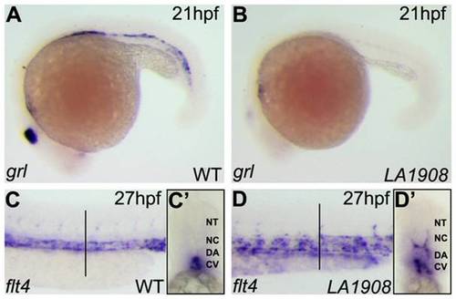
Aberrant expression of arterial and venous endothelial cell markers in LA1908 mutant embryos. A–B, Expression of grl, an arterial EC marker, was dramatically reduced in LA1908 mutant embryos (B versus A) at 21 hpf. C–D, Expression of the venous marker, flt4, was increased and localized ectopically in LA1908 mutant embryos at 27 hpf (D versus C). Vertical black bars, approximate location of vibratome cross-sections shown in C′–D′. C′–D′, Vibratome cross-section of embryos shown in C, D, revealing flt4 expression was appropriately restricted to the CV in 27 hpf wildtype embryos (C′), but is ectopically expressed in the DA and ISVs in LA1908 mutant embryos (D′). Embryos shown are representative from triplicate experiments performed on a minimum of 5 embryos per condition. |
| Genes: | |
|---|---|
| Fish: | |
| Anatomical Terms: | |
| Stage Range: | 20-25 somites to Prim-5 |