
Fig. 9
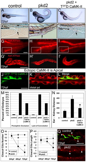
Constitutively active (T287D) CaMK-II reverses pronephric developmental defects. (A-F) Lateral views of 72 hpf entire embryos (A-C) and the cloaca (D-F; arrows indicate the end of the cloaca) after injection of pkd2 MO (4 ng) with or without T287D CaMK-II cDNA (30 pg). (G-I) Immunostaining α1 Na+/K+-ATPase identifies the pronephric ducts. (G2-I2) Z-stack renderings of the regions outlined by white boxes in G-I. (J-L) T287D CaMK-II localizes to apical surfaces of pronephric epithelial cells. (M) Quantification of the reversal of the pkd2 morphant phenotype (posterior: occlusion and anterior: convolution) by T287D CaMK-II (n=125-268). (N) Recovery of anterior migration of pronephric epithelial cells by ectopic T287D CaMK-II in pkd2 morphants as assessed by a decrease in posterior ear-anterior kidney distance (in μm; n=50-60). (O,P) Cloacal cilia number and length were determined at 24, 48 and 72 hpf. *P<0.005. (Q,R) Cloacal cilia disassemble but basal bodies remain apical as assessed by co-staining with acetylated tubulin (green) and γ-tubulin (red) at 72 hpf. Scale bars: 500 μm in A; 100 μm in D; 20 μm in G,G2 10 μm in J; 5 μm in Q. |
| Fish: | |
|---|---|
| Knockdown Reagent: | |
| Observed In: | |
| Stage Range: | Long-pec to Protruding-mouth |