
Fig. S1
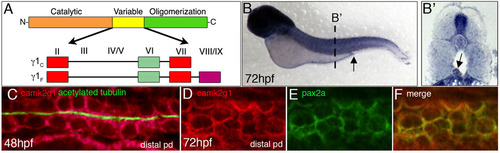
camk2g1 mRNA is expressed in the pronephric kidney. (A) Two alternative splice variants of the γ1 CaMK-II gene (γ1C and γ1F) were identified when cDNA from dissected kidneys or flow sorted kidney cells at 48 and 72 hpf was used as the template for RT-PCR. Alternative exon use in the variable domain is based on previous naming conventions. (B,B2) Whole-mount in situ hybridization was conducted using a γ1C probe at 72 hpf and is shown in whole mounts and in cryosectioned embryos. The cloaca is indicated by an arrow in B and the pronephric duct by an arrow in B2. (C) FISH was performed on 48 hpf embryos with the γ1C probe followed by immunostaining with acetylated tubulin to label pronephric ductal cilia. (D-F) Double fluorescent in situ hybridization demonstrates that γ1C CaMK-II and the pronephric kidney marker pax2a are co-expressed in the distal pronephros. |
| Genes: | |
|---|---|
| Antibody: | |
| Fish: | |
| Anatomical Terms: | |
| Stage Range: | Long-pec to Protruding-mouth |