
Fig. 5
- ID
- ZDB-FIG-110720-21
- Publication
- Banerjee et al., 2011 - A novel role for MuSK and non-canonical Wnt signaling during segmental neural crest cell migration
- Other Figures
- All Figure Page
- Back to All Figure Page
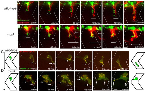
MuSK regulates maintenance of segmental neural crest migration. (A,B) Still images from time-lapse movies showing neural crest cells expressing membrane bound RFP [red, Tg(sox10:mRFP)vu234] and motor axons expressing GFP (green, Tg(mnx1:GFP)ml2) in wild-type (A) and musk mutant embryos (B). In musk mutants, neural crest cells migrate over a wider area of the somite (compare brackets between A and B). Mutant neural crest cells frequently form two streams, one migrating in close proximity to the motor axons, and one straying away from the central path towards the somite boundary (arrow). Scale bar: 10 μm. (C,D) Wild-type (C) and musk mutant (D) embryos expressing bioprobes for stable F-actin (mCherry-UtrCH) and total F-actin (Lifeact-GFP) in individual neural crest cells. A strong F-actin signal is visible at the leading front (arrow) of wild-type (C) and musk mutant (D) neural crest cells. Asterisks indicate filopodial projections towards somite boundaries (at 18 minutes in C; at 60, 206 and 236 minutes in D). Filopodial protrusions near the somite boundary are stabilized in the musk mutant embryo (marked by asterisk in D at 60, 206 and 236 minutes). Dashed lines indicate approximate positions of somite boundaries. Scale bars: 10 μm. See also Movies 2, 3, 4 and 5 in the supplementary material. |