
Fig. 4
- ID
- ZDB-FIG-110720-20
- Publication
- Banerjee et al., 2011 - A novel role for MuSK and non-canonical Wnt signaling during segmental neural crest cell migration
- Other Figures
- All Figure Page
- Back to All Figure Page
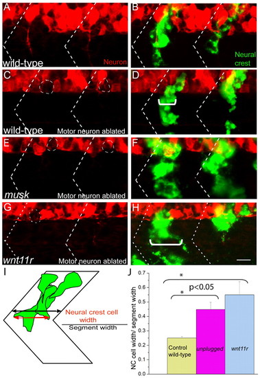
MuSK and Wnt11r signaling influences neural crest cell migration independent of motor axons. (A-D) In wild-type zebrafish embryos in which motoneurons were ablated (C,D), segmental neural crest cell migration is indistinguishable from that of wild-type embryos with intact motoneurons (A,B). (E-H) Neural crest cells migrate through a wider region of the somite (marked with white bracket) in motoneuron ablated musk (F) and wnt11r (H) mutant embryos, compared with the motoneuron-ablated wild-type control (C). Dashed circles in C, E and G indicate approximate positions of ablated motoneuron cell bodies. Dashed lines in A-H indicate approximate positions of somite boundaries. (I,J) Quantification of the neural crest cell migration phenotype in wild type, musk and wnt11r mutants was carried out by calculating the ratio of neural crest cell width (red arrow) with respect to segment width (black arrow; for details see Materials and methods). A minimum of five segments in which motoneurons were ablated were scored for each genotype, averaged and plotted with s.e.m. Scale bar: 10 μm. |