
Fig. s1
- ID
- ZDB-FIG-110624-37
- Publication
- Rothenaigner et al., 2011 - Clonal analysis by distinct viral vectors identifies bona fide neural stem cells in the adult zebrafish telencephalon and characterizes their division properties and fate
- Other Figures
- All Figure Page
- Back to All Figure Page
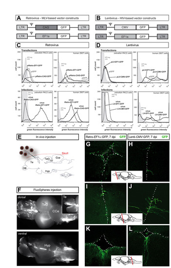
Viral transduction of zebrafish cells in culture and through in vivo injections into the ventricle of the adult zebrafish brain. (A,B) Retroviral MLV-based constructs (CAG or EF1α promoter) and lentiviral HIV-based constructs (CMV or EF1α promoter) used in this study. All self-inactivating viral vectors possessed an internal promoter to drive gfp expression. Ψ indicates retroviral packaging signal. (C,D) Flow-cytometry analysis (histograms) of GFP production after transfection (upper panels) and infection (lower panels) with retro- (C) and lenti- (D) viral vectors in zebrafish PAC2 cells (left panels) and (as control) human 293T cells (right panels). In all analyses, untransduced cells were monitored as control. In transfection experiments, the plasmid pCS2:gfp (pGFP) was used as control. This analysis identifies the retrovirus EF1α:GFP and the lentivirus CMV:GFP as the best combinations of promoters and virus types in zebrafish. (E) Schematic drawing (sagittal view) of the injection site into the telencephalic brain ventricle of the adult zebrafish brain. Solutions were injected anterior to the epiphysis. (F) Control injections with FluoSphere beads showed that the injected solution spreads throughout the ventricular region dorsal and ventral views of the brain at two days post-infection (dpi). (G-L) GFP expression in brains infected with Retro-EF1α:GFP (G,I,K) and Lenti-CMV:GFP (H,J,L) at 7 dpi (confocal stack projections, cross-sections at the levels indicated). The VZ is depicted with white dots. GFP expression could be detected in all ventricular areas, including telencephalon (G,H), mesencephalon (I,J) and medulla oblongata (K,L). Cce, corpus cerebellis; Hyp, hypothalamus; OB, olfactory bulb; Te, telencephalon; TeO, optic tectum; Val, Valvula. |