
Fig. 2
- ID
- ZDB-FIG-110624-32
- Publication
- Rothenaigner et al., 2011 - Clonal analysis by distinct viral vectors identifies bona fide neural stem cells in the adult zebrafish telencephalon and characterizes their division properties and fate
- Other Figures
- All Figure Page
- Back to All Figure Page
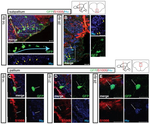
Maturation of GFP-positive cells into newborn neurons in the subpallial and pallial regions. Maturation was followed in the subpallium (upper panels) and pallium (lower panels) at various time points after infection (14, 21 and 28 dpi) (single optical sections from Retro-EF1α:GFP-injected brains, immunostained for GFP, S100β and/or HuC/D, white bars indicate the VZ). (A,B) In the subpallium, the majority of GFP-positive cells express HuC/D at 14 dpi (A, yellow arrows). Some labeled cells seem to move away from the VZ in chain-like assemblies (blue arrow). At 28 dpi, newborn neurons (yellow arrows) are small and exhibit short processes. Single-channel views are magnifications of the boxed areas. In B, all three cells indicated are HuC/D positive. (C-E) In the pallium, newborn HuC/D-positive neurons (white arrows) are still located close to the VZ at 28 dpi. Scale bars: 10 μm. |