
Fig. 5
- ID
- ZDB-FIG-110622-102
- Publication
- Nevin et al., 2011 - Topoisomerase II{beta} is required for lamina-specific targeting of retinal ganglion cell axons and dendrites
- Other Figures
- All Figure Page
- Back to All Figure Page
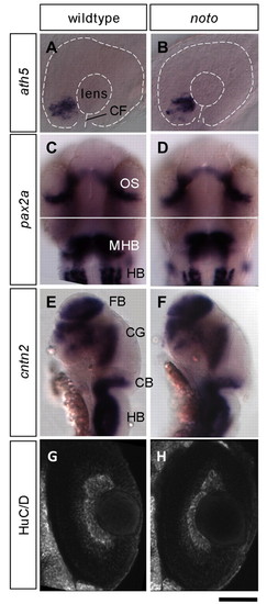
Expression patterns of early brain and eye development are preserved in noto mutants. (A,B) In situ hybridization with ath5 antisense probe at 28 hpf shows normal progression of neurogenesis in the wild-type and mutant zebrafish retina. Border of eye, lens and position of the choroid fissure (CF) are indicated by dashed lines. (C,D) pax2a expression at 28 hpf shows grossly normal development of the optic stalk (OS), midbrain-hindbrain boundary (MHB) and neurons of the hindbrain (HB) in both wild type and mutant. (E,F) cntn2 expression at 36 hpf shows grossly normal development of the forebrain (FB), several cranial ganglia (CG), the cerebellum (CB) and hindbrain (HB) in both wild type and mutant. (G,H) Confocal images of whole-mount HuC/D antibody-stained 48 hpf embryos. A similar population of HuC/D labeled ganglion cells (GCs) has developed in wild type (G) and mutant (H). Scale bar: 40 μm for A,B; 100 μm for C-F; 30 μm for G,H. |
| Genes: | |
|---|---|
| Fish: | |
| Anatomical Terms: | |
| Stage Range: | Prim-5 to Prim-25 |