
Fig. 2
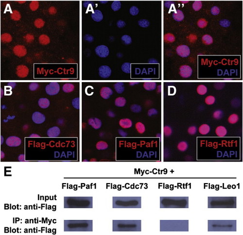
Fig. 2. Zebrafish PAF1C components localize to the nucleus and interact with each other in vivo. (A–A3) Panel (A3) shows the colocalization of Myc-tagged Ctr9 protein (red, A) and nuclear DAPI staining (blue, A2). (B) Flag-tagged Cdc73 (red) and DAPI staining (blue) colocalize in the nucleus. (C) Flag-tagged Paf1 (red) and DAPI staining (blue) colocalize in the nucleus. (D) Flag-tagged Rtf1 (red) and DAPI staining (blue) colocalize in the nucleus. (E) Flag-Paf1, Flag-Cdc73, and Flag-Leo1 coimmunoprecipitate with Myc-Ctr9. Input row shows 1 embryo equivalent of total protein lysate. IP row shows immunoprecipitated protein from 20 embryo equivalents of total protein lysate. |
Reprinted from Developmental Biology, 353(1), Langenbacher, A.D., Nguyen, C.T., Cavanaugh, A.M., Huang, J., Lu, F., and Chen, J.N., The PAF1 complex differentially regulates cardiomyocyte specification, 19-28, Copyright (2011) with permission from Elsevier. Full text @ Dev. Biol.