
|
Fig. 2
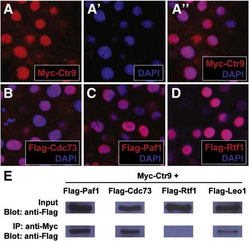
Fig. 2. Zebrafish PAF1C components localize to the nucleus and interact with each other in vivo. (A–A3) Panel (A3) shows the colocalization of Myc-tagged Ctr9 protein (red, A) and nuclear DAPI staining (blue, A2). (B) Flag-tagged Cdc73 (red) and DAPI staining (blue) colocalize in the nucleus. (C) Flag-tagged Paf1 (red) and DAPI staining (blue) colocalize in the nucleus. (D) Flag-tagged Rtf1 (red) and DAPI staining (blue) colocalize in the nucleus. (E) Flag-Paf1, Flag-Cdc73, and Flag-Leo1 coimmunoprecipitate with Myc-Ctr9. Input row shows 1 embryo equivalent of total protein lysate. IP row shows immunoprecipitated protein from 20 embryo equivalents of total protein lysate.
Reprinted from Developmental Biology, 353(1), Langenbacher, A.D., Nguyen, C.T., Cavanaugh, A.M., Huang, J., Lu, F., and Chen, J.N., The PAF1 complex differentially regulates cardiomyocyte specification, 19-28, Copyright (2011) with permission from Elsevier. Full text @ Dev. Biol.