
Fig. 6
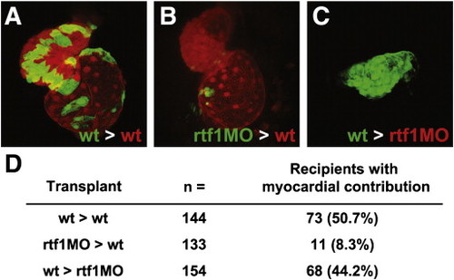
Fig. 6. Rtf1 regulates cardiomyocyte specification cell autonomously. (A–C) Confocal projections showing a ventral view of the heart in two day-old transplanted embryos. Wild type donor cells contribute robustly to the heart when transplanted into wild type recipients (A), while rtf1 morphant cells exhibit limited contribution when transplanted into a wild type recipient (B). Wild type cells are also able to generate myocardial tissue when transplanted into an rtf1 morphant host. (D) Summary of the results from transplantation experiments showing the contribution of donor cells to the myocardium of recipient embryos. |
Reprinted from Developmental Biology, 353(1), Langenbacher, A.D., Nguyen, C.T., Cavanaugh, A.M., Huang, J., Lu, F., and Chen, J.N., The PAF1 complex differentially regulates cardiomyocyte specification, 19-28, Copyright (2011) with permission from Elsevier. Full text @ Dev. Biol.