|
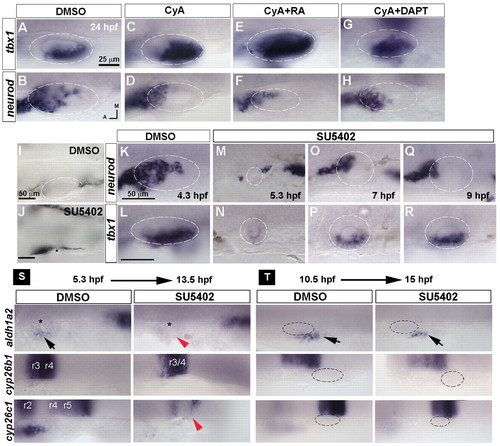
Temporal requirement for Hh and Fgf signals in otic development. (A-H) Expression of tbx1 and neurod in 24 hpf zebrafish embryos treated with DMSO, the Hh inhibitor cyclopamine A (CyA), CyA+RA and CyA+DAPT starting from 10.5 hpf. (I,J) Bright-field images of 30 hpf embryos treated with DMSO or the Fgf inhibitor SU5402 starting from 4.3 hpf reveals a requirement for Fgf signaling at this stage for otic placode induction. Asterisk indicates putative position of otic vesicle. (K-R) Stage-dependent influence of Fgf signaling on neurod and tbx1 expression. (S,T) In situ hybridization for aldh1a2, cyp26b1 and cyp26c1 in embryos treated with DMSO or SU5402 starting from 5.3 hpf or 10.5 hpf. Black arrows indicate mesodermal expression of aldh1a2; red arrowheads indicate suppression of aldh1a2 and cyp26c1 expression; asterisk indicates position of the otic placode. A-H, I,J and K-R are at the same magnification.
|