|
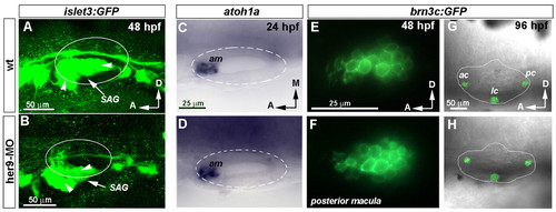
Depletion of her9 distorts the development of the SAG but not the generation of hair cells. (A,B) Three-dimensional reconstruction of the SAG at 48 hpf visualized in wild-type and her9-MO-injected zebrafish embryos carrying the islet3:GFP transgene. Arrowheads point to SAG branches. (C,D) Expression of atoh1a in wild-type and Her9 morphant embryos at 24 hpf. (E,F) Sagittal sections of posterior macula showing GFP fluorescence under the control of brn3c (pou4f3) reporter in a wild-type and her9-MO-injected embryo. (G,H) Three-dimensional reconstruction of selected confocal sections showing the otic cristae in wild-type and her9-MO-injected embryos. GFP in adjacent neuromasts of the lateral line has been removed for clarity. C,D, E,F and G,H are at the same magnification. SAG, statoacoustic ganglion; am, anterior macula; ac, anterior crista; lc, lateral crista; pc, posterior crista.
|