
Fig. 3
- ID
- ZDB-FIG-110128-18
- Publication
- Kinkhabwala et al., 2011 - A structural and functional ground plan for neurons in the hindbrain of zebrafish
- Other Figures
- All Figure Page
- Back to All Figure Page
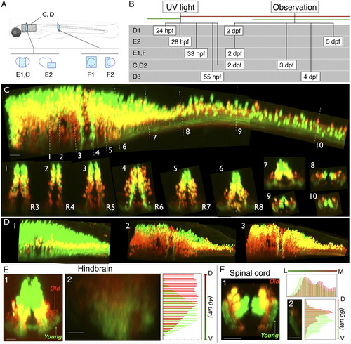
Age-related patterning within the medial glutamatergic, alx hindbrain stripe. (A) Location of images. (B) Timing of experiments. (C) Lateral view of alx:Kaede expression in a photoconverted fish shown from hindbrain through to spinal cord. White dashed lines indicate the rostrocaudal locations of the numbered cross-sections from different rhombomeres (R3–R8) and into spinal cord. (D, panels 1–3) Lateral views of hindbrain/spinal cord regions of alx:Kaede transgenic fish photoconverted/imaged at different times (timing shown in B). In this example we assigned colors so that green cells on the figure had no red in them at the imaging time, and neurons colored red on the figure are those with any red staining (they might also have had green but it was removed to make the youngest, pure green cells obvious). The yellow color here thus does not represent colabeling, but instead red and green neurons that overlap in the z direction. (E, panel 1) Reconstructed confocal cross-section shows that neuropil for older neurons (red) in hindbrain tends to be dorsal to that for younger neurons (green). (E, panel 2) Quantification of relative red/green expression at different dorsoventral locations within a cross-section of neuropil in rhombomere 7 from a different fish than E, panel 1; ventral is at the bottom. Red expression is shifted dorsally relative to green. (F) Photoconversion of an alx:Kaede transgenic fish imaged in spinal cord also shows age-related separation in the neuropil. In F, panel 1, a reconstructed confocal cross-section shows that neuropil for older neurons (red) tends to be medial and dorsal to that of younger neurons (green). (F, panel 2) Quantification of the dorsoventral distribution of red and green expression in the neuropil. (Scale bars, panel 2 in E and F, 10 μm; all other images, 20 μm.) |