
Fig. 1
- ID
- ZDB-FIG-110121-47
- Publication
- Zigman et al., 2011 - Zebrafish Neural Tube Morphogenesis Requires Scribble-Dependent Oriented Cell Divisions
- Other Figures
- All Figure Page
- Back to All Figure Page
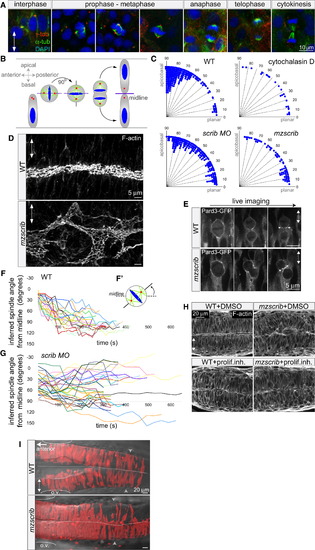
Scrib Regulates Mitotic Spindle Orientation and Neural Tube Morphogenesis (A and B) Immunostainings of neural keel progenitors (A) showing that the orientation of the mitotic spindle changes over the course of mitosis. γ-tubulin (centrosomes) is shown in red, α-tubulin (spindle) in green, and DAPI (DNA) in blue. This process results in the bilateral distribution of daughter cells as schematized in (B). (C) Quantification of mitosis orientation at anaphase. Chi-square analysis shows that the three distributions shown are highly significantly different from wild-type (WT) (n = 269): 3 μg/ml cytochalasin D-treated (n = 28, χ2 = 51, 1 degree of freedom [df]; p < 0.001), scrib morphant (n = 235, χ2 = 306, 8 df; p < 0.001), mzscrib mutant (n = 76, χ2 = 201, 2 df; p < 0.001). Although all three distributions are more homogeneous than WT, only cytochalasin D treatment results in a statistically randomized distribution (χ2 = 2.7, 3 df; p = 0.44). (D) Posterior hindbrain lumen morphology, defined by apical F-actin, with an abnormal, branched organization in the mzscrib mutant compared to WT. (E) Pard3-GFP localization to subapical foci upon cytokinesis (arrows) occurs normally in WT and misoriented mzscrib mutant neural keel progenitors. (F and G) Representation of mitotic spindle rotation in live WT (F; n = 20) and aberrantly rotating scrib morphant (G; n = 32) progenitors. Plots present the angle between inferred mitotic spindle axis and the midline over time. (F′) Scheme of spindle rotation in a mitotic progenitor. Mitotic spindle axis is shown as a solid line; midline is shown as a dashed line. (H) Inhibition of cell proliferation results in diminished neural tube morphogenesis defects in mzscrib embryos. (I) Requirement of Scrib for cross-midline cell divisions in the neural keel. Labeled cells in a 22 hours postfertilization (hpf) WT embryo have a bilateral distribution, but mzscrib mutant embryos have a predominantly unilateral distribution in the posterior hindbrain/anterior spinal cord region. Arrowheads indicate the position of the first somite; ovals mark the otic vesicle (o.v.); dotted line indicates the midline. In all panels, anterior is to the left and two-way arrows indicate the apicobasal axis of neuroepithelial progenitors. See also Figure S1. |
| Fish: | |
|---|---|
| Knockdown Reagent: | |
| Observed In: | |
| Stage Range: | 5-9 somites to 26+ somites |