
Fig. 2
- ID
- ZDB-FIG-110112-2
- Publication
- Robert-Moreno et al., 2010 - Characterization of new otic enhancers of the pou3f4 gene reveal distinct signaling pathway regulation and spatio-temporal patterns
- Other Figures
- All Figure Page
- Back to All Figure Page
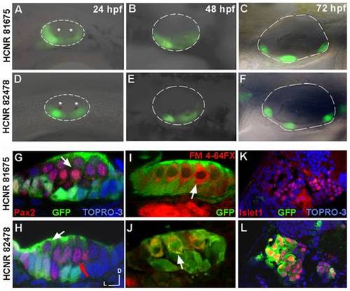
Spatial-temporal expression pattern of pou3f4 enhancers in the inner ear. (A–F) Lateral views of inner ears from zebrafish transgenic embryos for HCNR 81675 (A–C) and HCNR 82478 (D–F) enhancers analysed from 24 hpf to 72 hpf. In HCNR 81675 embryos at 24 hpf, GFP is observed in two broad domains comprising the sensory territories as observed by the otolith deposition (stars) (B). In HCNR 82478, GFP is already restricted to the anterior and posterior sensory macula from its onset as observed by GFP fluorescence relative to the otolith position (star). (C and F) GFP is found in the three sensory crista in 3-day old embryos in both transgenic zebrafish lines. Orientation is anterior (left) and dorsal (up). (G and H) Confocal transverse images of inner ear sensory patches immunostained with the anti-Pax2 antibody in 72 hpf embryos. In HCNR 81675 embryos, GFP is found in supporting cells but absent in hair-cells (Pax2 positive cells; pointed by a white arrow) (G). In contrast, HCNR 82478 embryos displayed GFP in supporting cells but also in hair-cells at lower levels (white arrow) whereas other hair-cells where completely devoid of GFP expression (red arrow). (I and J) Transverse confocal images of sensory patches of both enhancer embryos immunostained for GFP after the injection of the hair cell specific labelling marker FM 4-64FX. The same result was obtained in this experiment. (I) GFP is devoid in FM 4-64FX stained hair-cells in HCNR 81675 embryos (white arrow), whereas some hair-cells displayed GFP in HCNR 82478 embryos (J; white arrow). (K and L) Confocal images taken from the transverse section anterior to the first section from the otic vesicle. Co-immunostaining for anti-GFP and anti-islet1 protein reveals that only in HCNR 82478 transgenic embryos GFP is activated in the otic ganglion (L). (G–L) Lateral (left) and dorsal (up). |
| Gene: | |
|---|---|
| Antibody: | |
| Fish: | |
| Anatomical Terms: | |
| Stage Range: | Prim-5 to Protruding-mouth |