
Fig. 1
- ID
- ZDB-FIG-110112-1
- Publication
- Robert-Moreno et al., 2010 - Characterization of new otic enhancers of the pou3f4 gene reveal distinct signaling pathway regulation and spatio-temporal patterns
- Other Figures
- All Figure Page
- Back to All Figure Page
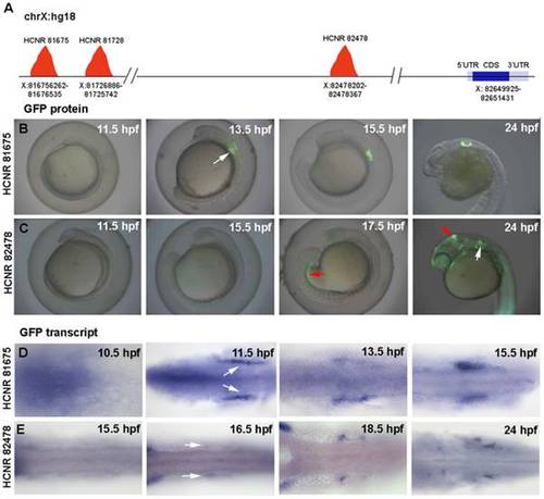
Temporal expression pattern of GFP driven by pou3f4 HCNR 81675 and HCRN 82478 enhancers. (A) Schematic representation of the POU3F4 locus in the human chromosome X (hg18 alignment) showing the position of the different inner ear enhancers relative to the POU3F4 coding sequence. (B–C) Onset of GFP protein expression in HCNR 81675 (B) and HCNR 82478 (C) transgenic embryos. Expression in the otic territory occurs at 13.5 hpf in HCNR 81675 and at 18.5 hpf in HCNR 82478 zebrafish embryos (white arrows), GFP in mesonephros and midbrain-hindbrain boundary (red arrows) is also detected in HCNR 82478 embryos. (D–E) Dorsal views of HCNR 81675 (D) and HCNR 82478 (E) transgenic embryos assayed by in-situ hybridization for GFP mRNA expression. In both cases GFP mRNA was detected in the otic field 2 hours before GFP protein was found (B–C). Orientation of the embryos is anterior (left) to posterior (right). |
| Gene: | |
|---|---|
| Fish: | |
| Anatomical Terms: | |
| Stage Range: | 1-4 somites to Prim-5 |