
FIGURE
Fig. S2
Fig. S2
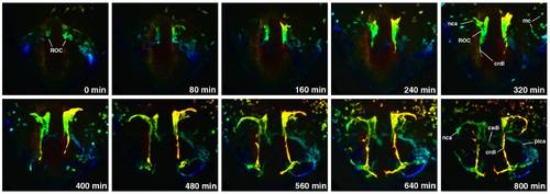
Depth-coded time-lapse images of cranial vessel formation in etsrp:GFP transgenic line from the 10-somite to approximately 22 hpf stages. Blue colors corresponded to the deepest cells while red colors mark the closest ones. Dorso-anterior view of the ROC region, anterior is to the top. This view is more rostral, compared to Fig. 5. |
Expression Data
Expression Detail
Antibody Labeling
Phenotype Data
Phenotype Detail
Acknowledgments
This image is the copyrighted work of the attributed author or publisher, and
ZFIN has permission only to display this image to its users.
Additional permissions should be obtained from the applicable author or publisher of the image.
Reprinted from Developmental Biology, 348(1), Proulx, K., Lu, A., and Sumanas, S., Cranial vasculature in zebrafish forms by angioblast cluster-derived angiogenesis, 34-46, Copyright (2010) with permission from Elsevier. Full text @ Dev. Biol.