
FIGURE
Fig. 4
Fig. 4
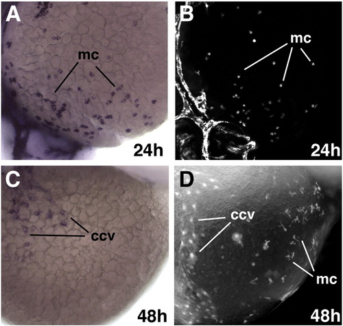
GFP protein is present in the myeloid cells after RNA is no longer transcribed in etsrp:GFP transgenic embryos. (A,C) In situ hybridization for GFP RNA at the ventral part of the yolk at 24 hpf (A) and 48 hpf (C). (B,D) GFP fluorescence at 24 hpf (B) and 48 hpf (D). Note that both GFP RNA and protein are present in the myeloid cells (mc) at 24 hpf while only GFP protein but not RNA is detected at 48 hpf. GFP RNA and protein are both present within the common cardinal vein (ccv) (C,D). |
Expression Data
Expression Detail
Antibody Labeling
Phenotype Data
Phenotype Detail
Acknowledgments
This image is the copyrighted work of the attributed author or publisher, and
ZFIN has permission only to display this image to its users.
Additional permissions should be obtained from the applicable author or publisher of the image.
Reprinted from Developmental Biology, 348(1), Proulx, K., Lu, A., and Sumanas, S., Cranial vasculature in zebrafish forms by angioblast cluster-derived angiogenesis, 34-46, Copyright (2010) with permission from Elsevier. Full text @ Dev. Biol.