
Fig. 3
- ID
- ZDB-FIG-101208-31
- Publication
- Yasuda et al., 2010 - Transgenic zebrafish reveals novel mechanisms of translational control of cyclin B1 mRNA in oocytes
- Other Figures
- All Figure Page
- Back to All Figure Page
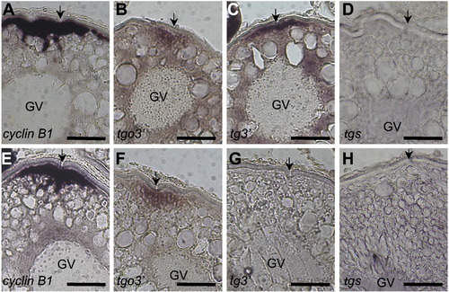
Localization of cyclin B1 and reporter mRNAs during oogenesis. (A–H) Section in situ hybridization using cyclin B1 (A, E) and gfp (B–D, F–H) probes of stage III (A–D) and stage IV (E–H) oocytes derived from wild-type (A, E) and transgenic fish carrying tgo3′ (B, F), tg3′ (C, G) or tgs (D, H) gene. Arrows indicate the micro-pile existing at the animal pole of fish oocytes. cyclin B1 and tgo3′ mRNAs were localized in the cortical cytoplasm beneath the animal pole of stage III (A, B) and IV (E, F) oocytes. tg3′ mRNA was localized to the animal pole of stage III oocytes (C) but disappeared in stage IV oocytes (G). tgs mRNA showed no localization during oogenesis (D, H). GV, germinal vesicle. Bars indicate 100 μm. |
Reprinted from Developmental Biology, 348(1), Yasuda, K., Kotani, T., Ota, R., and Yamashita, M., Transgenic zebrafish reveals novel mechanisms of translational control of cyclin B1 mRNA in oocytes, 76-86, Copyright (2010) with permission from Elsevier. Full text @ Dev. Biol.