
Fig. 2
- ID
- ZDB-FIG-101208-30
- Publication
- Yasuda et al., 2010 - Transgenic zebrafish reveals novel mechanisms of translational control of cyclin B1 mRNA in oocytes
- Other Figures
- All Figure Page
- Back to All Figure Page
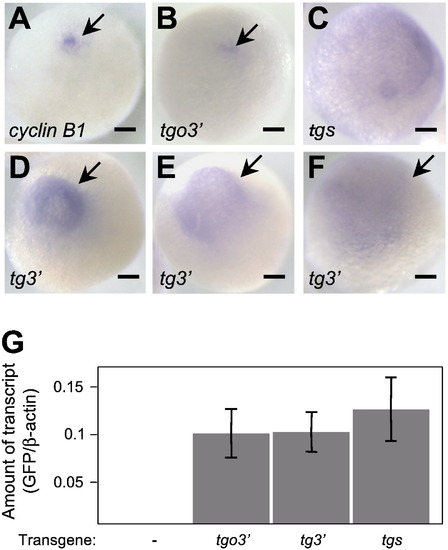
Localization of cyclin B1 and reporter mRNAs in full-grown oocytes. (A–F) Whole-mount in situ hybridization using cyclin B1 (A) and gfp (B–F) probes of full-grown oocytes derived from transgenic fish carrying the tgo3′ (A, B), tgs (C) or tg3′ (D–F) gene. Arrows indicate localized signals. cyclin B1 and tgo3′ mRNAs were localized as an aggregation (A, B), while tgs mRNA was distributed throughout the oocytes (C). tg3′ mRNA was distributed in the hemisphere of oocytes in various patterns, Classes I (D), II (E) and III (F) (see text). Bars indicate 100 μm. (G) Amount of the reporter mRNAs, normalized to that of β-actin mRNA. Real-time PCR using GFP primers showed that the transcripts of three reporter genes are deposited at similar levels in full-grown oocytes. Error bars indicate mean ± standard error of the mean (s.e.m.), n = 3. |
Reprinted from Developmental Biology, 348(1), Yasuda, K., Kotani, T., Ota, R., and Yamashita, M., Transgenic zebrafish reveals novel mechanisms of translational control of cyclin B1 mRNA in oocytes, 76-86, Copyright (2010) with permission from Elsevier. Full text @ Dev. Biol.