
Fig. 2
- ID
- ZDB-FIG-101123-5
- Publication
- Joseph, 2004 - Zebrafish IRX1b in the embryonic cardiac ventricle
- Other Figures
- All Figure Page
- Back to All Figure Page
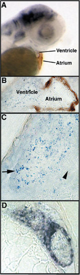
IRX1b expression in the ventricular chamber of the wild-type embryonic heart. A: Whole-mount in situ hybridization analysis of a wild-type embryo at 53 hours postfertilization (hpf) shows IRX1b expression (blue/purple color) in the ventricle. The atrium is stained by the S46 antibody (brown color), which recognizes an atrial specific myosin heavy chain isoform. B: A section of an IRX1b whole-mount in situ hybridized embryo at 53 hpf, with S46 antibody staining of the atrium, shows a “veil-like” IRX1b expression pattern in the ventricle. C: A sectioned embryo shows IRX1b expression predominantly in the trabeculated spongy myocardium of the ventricle at 53 hpf (blue/purple, arrow). The compact myocardial layer is indicated by an arrowhead. D:IRX1b expressing cells in the ventricle are arranged in a ring structure, as shown by the more intense staining in this section of a whole-mount in situ hybridized embryo at 54 hpf. |
| Gene: | |
|---|---|
| Antibody: | |
| Fish: | |
| Anatomical Terms: | |
| Stage: | Long-pec |