
- Title
-
Zebrafish IRX1b in the embryonic cardiac ventricle
- Authors
- Joseph, E.M.
- Source
- Full text @ Dev. Dyn.
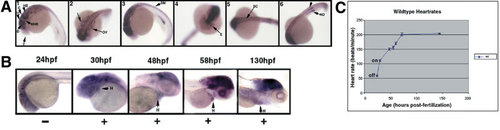
Zebrafish IRX1b gene expression at 1 and 2 days postfertilization (dpf). A: Panel 1: At 22 hours postfertilization (hpf), IRX1b is expressed in the central nervous system. A lateral view of a 26 somite stage embryo (22 hpf) with the anterior at the left and the posterior to the right. Dorsal is at the top and ventral is at the bottom. Expression is particularly strong in the midbrain and hindbrain structures (the diencephalon, tectum, and cerebellum) but is excluded from the forebrain and the midbrain–hindbrain junction. T, telencephalon; D, diencephalon; TC, tectum; MHB, midbrain–hindbrain junction; C, cerebellum; HB, hindbrain. Panel 2: A dorsal view of a 22 hpf embryo shows IRX1b expression in the otic vesicles (OV). Panel 3: At 24 hpf, IRX1b is expressed in the somitic mesoderm (SM). Panel 4: An anterior view of a 24 hpf embryo shows IRX1b expression also occurs in the eye (E). Panel 5: A dorsal view of a 24 hpf embryo shows expression in the spinal cord (SC) and somitic mesoderm. Panel 6: At 24 hpf, expression is also seen in the two nephric ducts (ND, arrow). B:IRX1b is not detected in the newly formed heart tube at 24 hpf (left-most panel). By 30 hpf, weak IRX1b expression is detected in the primary heart tube (H, heart). This expression continues at the later stages examined (48, 58, 130 hpf; all other panels). C: The onset of IRX1b cardiac expression is correlated with an increase in embryonic heart rate. The heart rates of wild-type embryos were counted at 24, 30, 48, 55, 60, 72, and 144 hpf. The average heart rates (beats per minute) were plotted versus age (hpf) with error bars indicating standard deviations. The greatest change in beats per minute per hour of ageing occurs between 24 and 30 hpf. EXPRESSION / LABELING:
|
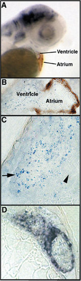
IRX1b expression in the ventricular chamber of the wild-type embryonic heart. A: Whole-mount in situ hybridization analysis of a wild-type embryo at 53 hours postfertilization (hpf) shows IRX1b expression (blue/purple color) in the ventricle. The atrium is stained by the S46 antibody (brown color), which recognizes an atrial specific myosin heavy chain isoform. B: A section of an IRX1b whole-mount in situ hybridized embryo at 53 hpf, with S46 antibody staining of the atrium, shows a “veil-like” IRX1b expression pattern in the ventricle. C: A sectioned embryo shows IRX1b expression predominantly in the trabeculated spongy myocardium of the ventricle at 53 hpf (blue/purple, arrow). The compact myocardial layer is indicated by an arrowhead. D:IRX1b expressing cells in the ventricle are arranged in a ring structure, as shown by the more intense staining in this section of a whole-mount in situ hybridized embryo at 54 hpf. EXPRESSION / LABELING:
|
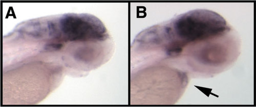
IRX1b gene expression in silent heart mutant embryos. A,B: Whole-mount in situ hybridization shows weakened IRX1b cardiac expression in the silent heart genetic mutant (tc300b allele, at 54 hours postfertilization [hpf]; compared with IRX1b expression in a wild-type sibling embryo at 54 hpf (B, heart indicated by arrow). EXPRESSION / LABELING:
|
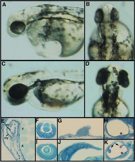
Phenotype of embryos injected with a morpholino designed to block IRX1b translation. A: Injection of an IRX1b morpholino (IRX1b-MO) antisense oligonucleotide prevents proper central nervous system development as shown here in these lateral views of a live 2 days postfertilization (dpf) embryo injected with a high dose of 1,000 μM. B: Dorsal view of the same IRX1b morpholino-injected embryo. C: A wild-type sibling of the morpholino-injected embryo shown in A and B. D: Dorsal view of the same wild-type embryo. E: IRX1b-MO–injected embryos develop with a distinctive ventricular (V) and atrial cardiac chamber (A); these chambers both have an endocardium (en) and a myocardium (my). F: Methylene blue azure II stained sections of IRX1b morpholino-injected embryos show other embryonic structures to be disrupted. The eye of IRX1b-MO–injected embryos has an altered retinal laminar pattern (compare with I). G: Fin outgrowth is reduced (compare with J). H: Semicircular canals do not form in the otic vesicles of IRX1b-MO–injected embryos, but otoliths do form (compare with K). I: Section through the eye of a wild-type sibling shows the normal laminar pattern of retinal neurons. J: Section through a wild-type fin shows the extent of fin outgrowth as well as the usual cell arrangement within the fin. K: Section through a wild-type sibling otic vesicle shows the presence of semicircular canal precursors (arrows). PHENOTYPE:
|