
Fig. 1
- ID
- ZDB-FIG-101123-4
- Publication
- Joseph, 2004 - Zebrafish IRX1b in the embryonic cardiac ventricle
- Other Figures
- All Figure Page
- Back to All Figure Page
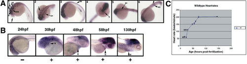
Zebrafish IRX1b gene expression at 1 and 2 days postfertilization (dpf). A: Panel 1: At 22 hours postfertilization (hpf), IRX1b is expressed in the central nervous system. A lateral view of a 26 somite stage embryo (22 hpf) with the anterior at the left and the posterior to the right. Dorsal is at the top and ventral is at the bottom. Expression is particularly strong in the midbrain and hindbrain structures (the diencephalon, tectum, and cerebellum) but is excluded from the forebrain and the midbrain–hindbrain junction. T, telencephalon; D, diencephalon; TC, tectum; MHB, midbrain–hindbrain junction; C, cerebellum; HB, hindbrain. Panel 2: A dorsal view of a 22 hpf embryo shows IRX1b expression in the otic vesicles (OV). Panel 3: At 24 hpf, IRX1b is expressed in the somitic mesoderm (SM). Panel 4: An anterior view of a 24 hpf embryo shows IRX1b expression also occurs in the eye (E). Panel 5: A dorsal view of a 24 hpf embryo shows expression in the spinal cord (SC) and somitic mesoderm. Panel 6: At 24 hpf, expression is also seen in the two nephric ducts (ND, arrow). B:IRX1b is not detected in the newly formed heart tube at 24 hpf (left-most panel). By 30 hpf, weak IRX1b expression is detected in the primary heart tube (H, heart). This expression continues at the later stages examined (48, 58, 130 hpf; all other panels). C: The onset of IRX1b cardiac expression is correlated with an increase in embryonic heart rate. The heart rates of wild-type embryos were counted at 24, 30, 48, 55, 60, 72, and 144 hpf. The average heart rates (beats per minute) were plotted versus age (hpf) with error bars indicating standard deviations. The greatest change in beats per minute per hour of ageing occurs between 24 and 30 hpf. |
| Gene: | |
|---|---|
| Fish: | |
| Anatomical Terms: | |
| Stage Range: | 26+ somites to Day 5 |