
Fig. S6
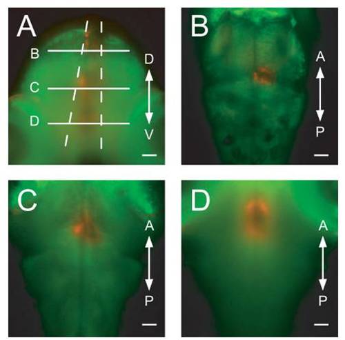
Confirmation of precise light application with optic fibers. A Gal4s1101t; UAS:Dendra-kras animal was imaged from two orthogonal directions, after Dendra had been converted with an optic fiber (50-μm diameter) placed over the dorsal surface of the animal. (A) Transverse view. The converted cone of red cells is visible. Dashed lines represent the theoretical divergence angle for 50-μmfibers and (B–D) indicate the positions of the optical sections in the panels (B–D). (B–D) Horizontal optical sections at the positions indicated in panel (A). D, dorsal, V, ventral, A, anterior, and P, posterior. (Scale bars, 50 μm.) |