
Fig. 4
- ID
- ZDB-FIG-101115-27
- Publication
- Qin et al., 2009 - Genetic evidence for shared mechanisms of epimorphic regeneration in zebrafish
- Other Figures
- All Figure Page
- Back to All Figure Page
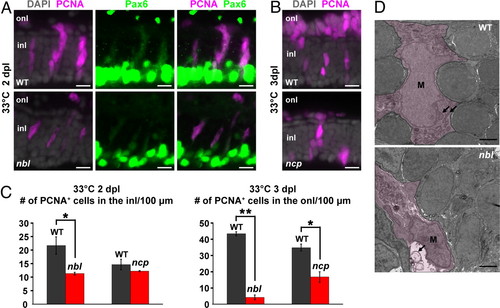
Retinal regeneration defects of nbl and ncp. (A) Neurogenic clusters at 2 dpl in the inner nuclear layer (inl) immunolabeled with anti-PCNA (magenta) and weakly labeled with anti-Pax6 (green) in WT and nbl. Note that Pax6 is also expressed at high levels in amacrine cells at the inner boundary of the inl. (B) PNCA+ photoreceptor progenitors at 3 dpl in the outer nuclear layer (onl) of WT and ncp. (C) Number of PNCA+ cells in the inl or onl per 100 μm of linear length retina at 2 or 3 dpl, respectively. Error bars represent SEM for 3 individuals. *, P < 0.05; **, P < 0.0001. (D) Transmission electron micrographs of injury-activated Müller glia in WT and nbl. See text for description of temperature shift paradigm. Müller glia (M) are shown by the magenta wash. Mitochondria (arrows) in Müller glia of WT appear normal after 8 h at 33°C, whereas in nbl mutants, Müller glia contain swollen mitochondria. (Scale bars: 10 μm in A and B; 100 μm in D.) |