
FIGURE
Fig. 1
- ID
- ZDB-FIG-101115-24
- Publication
- Qin et al., 2009 - Genetic evidence for shared mechanisms of epimorphic regeneration in zebrafish
- Other Figures
- All Figure Page
- Back to All Figure Page
Fig. 1
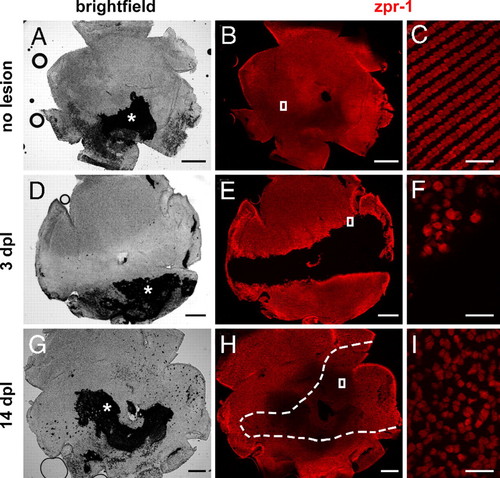
Cone photoreceptor regeneration in adult zebrafish. Flat-mounted zebrafish retinas are immunolabeled with cone-specific zpr-1 (red). Retinas are oriented dorsal up, ventral down, nasal left, and temporal right. (A, B) Intact retina. Asterisk, attached retinal pigment epithelium. (D, E) At 3 days after exposure to intense light, cones are missing in a horizontal band across the retina. (G, H) By 14 days, cones have regenerated within the lesioned region (dashed lines). (C, F, and I) Magnified images of the boxes in B, E, and H, respectively. (Scale bars: 300 μm in A, B, D, E, G, and H; 20 μm in C, F, and I.) |
Expression Data
Expression Detail
Antibody Labeling
Phenotype Data
Phenotype Detail
Acknowledgments
This image is the copyrighted work of the attributed author or publisher, and
ZFIN has permission only to display this image to its users.
Additional permissions should be obtained from the applicable author or publisher of the image.
Full text @ Proc. Natl. Acad. Sci. USA