
Fig. 3
- ID
- ZDB-FIG-101104-4
- Publication
- Raphael et al., 2010 - Schwann cells reposition a peripheral nerve to isolate it from postembryonic remodeling of its targets
- Other Figures
- All Figure Page
- Back to All Figure Page
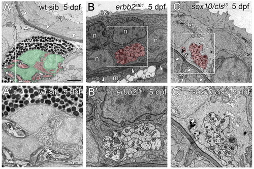
The PLLn is mislocalized in mutants lacking Schwann cells. (A) At 5 dpf, the PLLn of wild-type siblings has migrated from the epidermis (ep), across the basement membrane (arrowheads) into the muscle (m) (n=28). (B,C) By contrast, at 5 dpf in the Schwann cell-deficient mutants erbb2st61 (B) and sox10/clst3 (C), the PLLn remains in the epidermis, outside of the basement membrane (arrowheads, n=7 in B and n=6 in C). Defasciculation of the mutant nerves is also observed (asterisk, C). (A′,B′,C′) Higher magnification of boxed regions in A,B,C, respectively. Scale bars: 2 μm in A,B,C; 1 μm in A′,B′,C′. Schwann cells are pseudocolored green and axons red. Abbreviations: ep, epidermis; m, muscle; p, pigment; n, neuromast cell. |